The wrinkled outer layer of the brain is the cerebral cortex, and it is divided into four lobes. Underneath it sit the structures that handle memory, emotion, hormones and the automatic things your body does without being asked. The pictures below are anatomical drawings, not photographs.
Every structure is explained in plain text further down the page, so you never have to read anything off a picture. On the Outer Brain diagram you can also select a region to jump straight to its description.
Outer Brain
The four lobes of the cerebral cortex, and what sits behind them.

Frontal lobe
Where it is. The front of the brain, behind your forehead.
What it does
- Planning, decision-making and reasoning
- Holding something in mind while you work on it
- Impulse control
- Voluntary movement
- Much of what we mean by personality
How it develops. The frontal lobes are among the last areas of the brain to mature. They may not be fully developed until halfway through the third decade of life — the mid-to-late 20s.
Phineas Gage. In 1848 a railway worker named Phineas Gage survived an iron rod passing through his frontal lobe. The change in him afterwards is one of the first pieces of evidence that a specific part of the brain shapes judgment and behavior. The story is usually told as though he was ruined for life. He was not: re-examining the record, Macmillan and Lena found he eventually made a surprisingly good psychosocial adaptation to the injury, and argue that the structure his later work gave him helped him get there.
Parietal lobe
Where it is. Behind the frontal lobe, towards the top and back of the head.
What it does
- Touch, pressure, pain and temperature
- Knowing where your body is without looking
- Putting information from different senses together
Occipital lobe
Where it is. The back of the brain.
What it does
- Processing what the eyes send
- Recognizing shape, motion and color
Temporal lobe
Where it is. The sides of the brain, roughly behind the ears.
What it does
- Hearing
- Understanding language
- Forming and retrieving memories
- Recognizing objects and faces
Cerebellum
Where it is. At the back and underneath, behind the brain stem.
What it does
- Coordinating voluntary movement
- Balance and posture
- Fine motor control, like writing or catching a ball
- Motor learning — why a practiced movement becomes smooth
- Newer research also links the cerebellum to attention and emotional processing
Small compared with the rest of the brain, the cerebellum holds more than half of the neurons in your whole body.
Brain stem
Where it is. The stalk between the brain and the spinal cord.
What it does
- Carries every signal passing between the brain and the body
- Its three parts — the midbrain, the pons and the medulla — handle reflexes, relaying, and the automatic functions that keep you alive
Each of those three parts has its own entry below. The brain stem is the name for them together, not a fourth structure.
Inside the Brain
The midline structures, from the corpus callosum arching across the top to the brain stem leaving at the bottom.
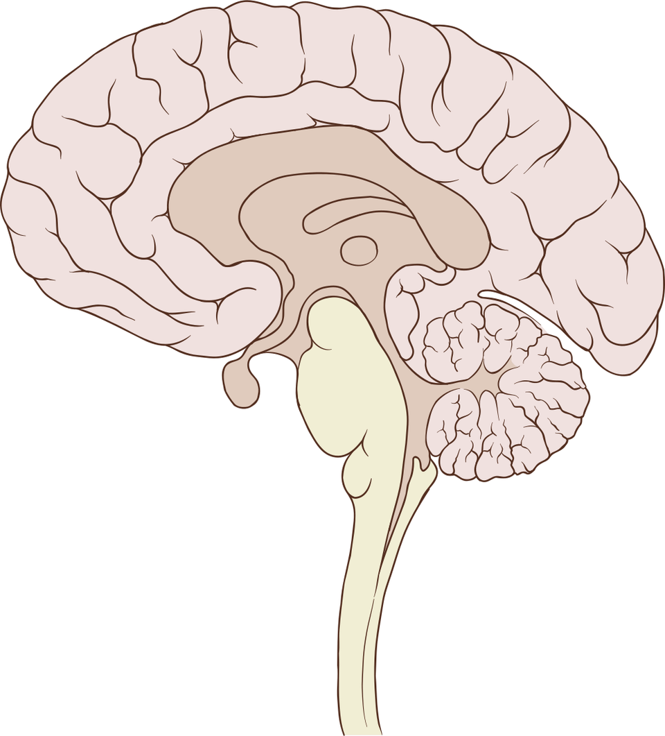
The brain cut straight down the midline and seen from the left. The folded cerebral cortex wraps the outside; the corpus callosum, the broad band of fibres joining the two hemispheres, arches across the centre; the deep midline structures lie below and behind it; and the brain stem descends at the back, with the cerebellum folded behind it.
The illustration carries no labels. Each midline structure is named, located and explained in the list below.

Corpus callosum
Where it is. A broad band across the midline, joining the two hemispheres.
What it does
- Carries nerve fibers between the left and right hemispheres
Fornix
Where it is. A curved bundle of fibers beneath the corpus callosum.
What it does
- Carries signals out of the hippocampus towards the mammillary bodies, as part of the limbic system
Thalamus
Where it is. Deep in the center of the brain, just above the brain stem.
What it does
- A relay station — nearly all sensory information passes through it on the way to the cortex
- Involved in alertness, sleep and consciousness
Hypothalamus
Where it is. Just below the thalamus, above the pituitary gland.
What it does
- Body temperature
- Hunger and thirst
- Setting the daily circadian rhythm, through its suprachiasmatic nucleus
- Releasing hormones, and instructing the pituitary gland
- Part of the body’s stress response
Pituitary gland
Where it is. A pea-sized gland hanging just below the hypothalamus.
What it does
- Releases hormones that affect growth, metabolism, the thyroid, the adrenal glands and the reproductive organs
- Takes its instructions from the hypothalamus
Pineal gland
Where it is. A small gland near the center of the brain, behind the thalamus.
What it does
- Makes melatonin
- Helps set the daily sleep and waking cycle
How it develops. Melatonin levels are high in childhood and fall around puberty. They are believed to influence the timing of sexual development.
Midbrain
Where it is. The top section of the brain stem, above the pons.
What it does
- Eye movement and focusing
- Reflex responses to sights and sounds, like turning your head towards a loud noise
- Passing visual and auditory information onward
- Alertness and arousal
- Contains the substantia nigra, which produces much of the brain’s dopamine
Pons
Where it is. The middle section of the brain stem, between the midbrain and the medulla.
What it does
- Relays signals between the cerebrum and the cerebellum
- Carries several of the cranial nerves
- Helps modulate breathing
- Generates REM sleep — the stage in which most dreaming happens
Medulla oblongata
Where it is. The lowest section of the brain stem, where it meets the spinal cord.
What it does
- Breathing
- Heart rate and blood pressure
- Swallowing, coughing and vomiting
- Relaying signals between the brain and the spinal cord
Spinal cord
Where it is. Runs from the base of the brain stem down inside the spine.
What it does
- Carries sensory information up to the brain
- Carries motor commands down to muscles and glands
- Handles some reflexes on its own, without involving the brain
Memory & Emotion
Deeper still — the amygdala and the hippocampus, set in the temporal lobes.
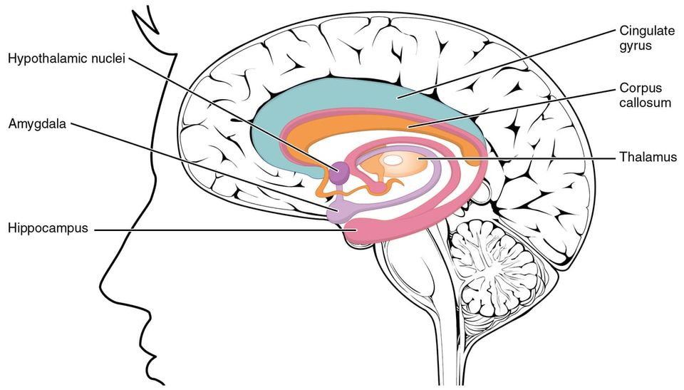
The brain seen from the midline, with the structures of the limbic system drawn in colour and labelled on the image. Arching over the centre are the cingulate gyrus (the outer band) and, just within it, the corpus callosum. The thalamus sits at the core, with the hypothalamic nuclei below it. Towards the front is the amygdala, and curving behind and below it, the hippocampus.
The amygdala and the hippocampus are drawn once here, on the midline, but there are really two of each — one deep in the left temporal lobe and one in the right. This diagram shows the limbic system as a whole; the two structures are described on their own below.

Amygdala
Where it is. Almond-shaped, one in each temporal lobe, part of the limbic system.
What it does
- Detecting threat
- Processing fear
- Attaching emotional weight to memories, which is why frightening events are remembered so vividly
- A central part of the body’s stress response
- Heavily implicated in anxiety and in PTSD
Hippocampus
Where it is. A curved structure deep inside each temporal lobe, just below and behind the amygdala.
What it does
- Forming new long-term memories
- Navigation
Sources
- Lenroot & Giedd (2010), Annual Review of Clinical Psychology (PMC2892678)
- The Teen Brain: 7 Things to Know — NIH National Institute of Mental Health
- Macmillan & Lena (2010), Rehabilitating Phineas Gage, Neuropsychological Rehabilitation (PMID 20480430)
- Brain Basics: Know Your Brain — NIH National Institute of Neurological Disorders and Stroke
- Neuroanatomy, Occipital Lobe — StatPearls, NIH National Library of Medicine
- Neuroanatomy, Temporal Lobe — StatPearls, NIH National Library of Medicine
- Neuroanatomy, Cerebellum — StatPearls, NIH National Library of Medicine
- Cerebellum: What It Is, Function & Anatomy — Cleveland Clinic
- Neuroanatomy, Brainstem — StatPearls, NIH National Library of Medicine
- Neuroanatomy, Corpus Callosum — StatPearls, NIH National Library of Medicine
- Neuroanatomy, Limbic System — StatPearls, NIH National Library of Medicine
- Neuroanatomy, Thalamus — StatPearls, NIH National Library of Medicine
- Neuroanatomy, Hypothalamus — StatPearls, NIH National Library of Medicine
- Physiology, Hypothalamus — StatPearls, NIH National Library of Medicine
- Physiology, Pituitary Gland — StatPearls, NIH National Library of Medicine
- Physiology, Pineal Gland — StatPearls, NIH National Library of Medicine
- Neuroanatomy, Mesencephalon (Midbrain) — StatPearls, NIH National Library of Medicine
- Neuroanatomy, Pons — StatPearls, NIH National Library of Medicine
- Physiology, REM Sleep — StatPearls, NIH National Library of Medicine
- Neuroanatomy, Medulla Oblongata — StatPearls, NIH National Library of Medicine
- Neuroanatomy, Spinal Cord — StatPearls, NIH National Library of Medicine
- Neuroanatomy, Amygdala — StatPearls, NIH National Library of Medicine
- Amygdala: What It Is and What It Controls — Cleveland Clinic
- Neuroanatomy, Hippocampus — StatPearls, NIH National Library of Medicine